The Brain Tumor Program provides an extensive referral system for diagnosis, management, rehabilitation and long-term treatment, and offers the latest advanced equipment delivering radiation therapy with never-before-attained precision.
“Since we’re located in an area that includes the highest incidence of brain tumor, developing this regional brain tumor program is very important,” says Dr. Edward Flotte, assistant professor of neurosurgery.
The program’s Brain Tumor Laboratory preserves samples of brain tumors resected, or surgically removed, at the Medical Center for genetic testing. Each specimen is correlated with a clinical database, which generally includes digitized magnetic resonance image files.
The program participates in several National Cancer Group programs and protocols that investigate new treatment, and it takes part in several multicenter trials of new chemotherapeutic agents. One study is examining why central and north Mississippi and west Tennessee record the highest rates of brain tumor cases nationally.
The basic science research focus is on the expression of developmental genes in brain tumors. Previous work has shown that genes that are required for the formation of neurons and glia in the embryo are also expressed in brain tumors. These genes have the potential to act as tumor markers to aid in diagnosis.
For example, in some cases it is difficult to distinguish between oligodendrogliomas and astrocytomas (two related tumor types) based on histology, or microscopic tissue studies, alone. The difference is clinically significant because oligodendrogliomas are more responsive to chemotherapy than astrocytomas.
As might be expected, genes involved in the production of oligodendrocytes (for example, Olig2) are expressed at higher levels in oligodendrogliomas than in astrocytomas, indicating that they may be clinically significant markers. Because the cell-of-origin remains unknown for virtually all brain tumors, by looking at the expression pattern of these lineage-determining genes doctors also may obtain clues as to where the tumors arise.
Besides advancing knowledge about specific types of brain tumors, having a regional brain tumor center in the state improves patient care for Mississippians, from diagnosis to rehabilitation.
“This is the first time all specialties are covered in one place for the treatment of brain tumors,” says Flotte, who coordinates the program along with Dr. James Fontanesi, chairman of the Department of Radiation Oncology, and Dr. Ruth Fredericks, clinical assistant professor of neurology. “Tumors of the brain are different from tumors in any other part of the body, because they must be resected without damaging any of the surrounding structures of the brain.”
Fontanesi says the foundation for the program’s ability to provide the highest quality service is built more on its multidisciplinary approach to patient care than its high-tech equipment.
“From the moment the patient walks in, everyone involved in the program—from the radiation oncologist to the neuro-oncologist to the neurosurgeon—cooperates to determine the best course of treatment for each individual,” Fontanesi says. “It’s much more comfortable for the patients to see all of us before their operation so that we can address all of their questions in the clinic.
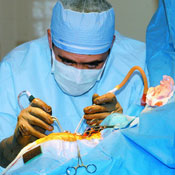 Dr. Edward Flotte resects a brain tumor using the intraoperative MRI, which allows him to ‘see’ the tumor while performing the procedure.
Once a possible tumor has been identified, the Department of Neurosurgery can use a number of methods for diagnosis, from open biopsy to frame-based and image-guided stereotactic biopsy to Interventional MRI-guided biopsy. Following the biopsy, a neuropathologist can perform a complete pathologic analysis to provide a more tailored approach to the management of some tumors.
The Department of Neurosurgery uses advanced technology to manage brain and spinal tumors—including frameless stereotaxy, intraoperative ultrasound, intraoperative cortical motor and language mapping and, in some cases, performing brain surgery while the patient is awake to minimize the risk of neurologic deficits after the operation. Degradable chemotherapy wafers or balloons containing radioactive liquid iodine may be placed directly in the surgical cavity after removal of the tumor.
The Medical Center was the third institution in the country to use the Intraoperative MRI, which allows surgeons to actually see tumors during surgery and resect them much more easily. But radiation therapy equipment that is just coming online has the Brain Tumor Program staff buzzing.
Medical Center neurosurgeons use the Gamma Knife machine at the Robert Smith Gamma Knife Center to perform radiosurgery—what has been called “brain surgery without a knife.” Radiosurgery delivers high-dose, highly focused radiation to a tumor to kill it while sparing the normal brain structures. Patients with small and deep-seated tumors are most likely to benefit from the radiosurgery. Unlike conventional radiation therapy, which can take weeks, radiosurgery is performed in one session as an outpatient procedure.
The Cancer Institute acquired a newer machine for performing radiosurgery—the Elekta Synergy—earlier this year. Unlike the Gamma Knife, which can only deliver stereotactic radiation to lesions in the head, the Elekta Synergy allows radiation oncologists to administer a precise dose of radiation in the brain, spine or anywhere else in the body.
“This linear accelerator significantly increases the magnitude of how precisely we can deliver the radiation,” Fontanesi says. “We are able to give a much higher dose in a much shorter amount of time.”
Additional treatment planning and therapy equipment is scheduled to be available in the Department of Radiation Oncology, located in the Cancer Institute, late this summer.
The Medical Center will be the first to receive the improved radiation source Californium-252, which has been shown to have advantages over traditional radiation, especially in brain tumors. By a procedure called brachytherapy, radiation oncologists can implant Californium-252 directly into the tumor.
“Californium-252 emits neutrons that are much more potent at killing cells, but you have to be very precise about where these neutrons go,” Fontanesi says. “We will be able to apply this substance by using a special temporary tube that the Californium can be slipped in and out of daily.”
“By placing ‘seeds’ of Californium directly into the tumor, the surrounding tumor will be radiated, leaving the remainder of the brain unharmed,” Flotte says. Between 30 percent and 50 percent of patients who formerly received radiation of the entire brain experienced dementia within 12 to 24 months of the procedure, he says.
The Medical Center will be one of the first 10 academic centers in the country to employ intensity-modulated radiation therapy, which uses new image-fusion software to combine multiple imaging sources such as CT, MRI, PET and SPECT to better define lesions.
The Medical Center also is working in conjunction with the National Aeronautics and Space Administration to develop a three-dimensional holographic system to improve treatment planning.
“This exciting technology will allow our neurosurgeons to view specific parts of the brain so they can figure out the best way to approach the tumor before they even begin the surgery,” Fontanesi says, adding that the most amazing aspect of the technological boom in the Brain Tumor Program is what it means for the patients. “Neurosurgeons are able to go into the brain and get to lesions where they have never been able to before. Chemotherapy agents are being applied more precisely than before. All with very few side effects for the patient.”
Flotte agrees that patients will see real-life benefits of the new technology.
“All this technology is geared to one thing—making sure each patient can have good quality of life,” he says. “Survival rates are still poor for the most common types of brain tumors, but this will gradually allow us to improve our surgical techniques, resect as much of the tumor as possible and ultimately extend the life expectancy and quality of life for our patients.”